You can listen to him here (in Polish).
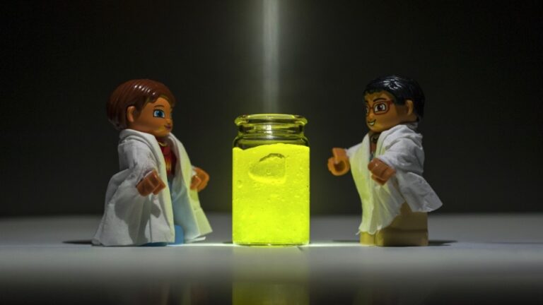
If we were talking about food, most experts would choose the former, but in the case of energy storage the opposite is true. It turns out that more energy can be stored by charging less often, but right up to 100%.
At least, this is the conclusion arrived at from research carried out by a team of scientists at the IPC PAS. Although the studies involved idealized two-dimensional lattice systems, at the end of the day, a principle is a principle. Dr. Anna Maciołek, one of the authors of the work published in Physical Review E, describes it as follows. “We wanted to examine how the manner in which energy is stored in a system changes when we pump energy in the form of heat into it, in other words – when we heat it locally.” It is known that in systems heat spreads out and diffuses. But is the collection of energy influenced by the way it is delivered; speaking professionally “the delivery alignment”? Does it matter whether we provide a lot of energy over a short period of time, none for a long time and then again a lot of energy, or small portions of energy one after the other, almost without any breaks?
Cyclic energy supply is very common in nature. We provide ourselves with energy in just this manner by eating. The same number of calories can be provided in one or two large portions eaten during the day, or broken down into 5-7 smaller meals with shorter breaks between them. Scientists are still arguing about which regimen is better for the body.
However, when it comes to two-dimensional lattice systems, it is already known that in terms of storage efficiency the “less often and a lot” method wins. “We noticed that the amount of energy the system can store varies depending on the portion size of the energy and the frequency of its provision. The greatest amount is when the energy portions are large, but the time intervals in between their supply are also long,” explains Yirui Zhang, a PhD student at the IPC PAS. “Interestingly, it turns out that if we divide this sort of storage system internally into compartments or indeed chambers, the amount of energy that can be stored in such a divided-up “battery”- if it were possible to construct – increases. In other words, three small batteries can store more energy than one large one,” says the researcher. All this, assuming that the total amount of energy put into the system remains the same, and only the method of its delivery changes.
Although the research carried out by the IPC PAS team is quite basic and simply shows the fundamental principle governing energy storage in magnets, its potential applications cannot be overestimated. Let’s imagine, for example, the possibility of charging an electric car battery not in a few hours, but in just under twenty minutes, or a significant increase in the capacity of such batteries without changing their volume, i.e. extending the range of the car after one charge. The new discovery may also, in the future, change the methods of charging different types of batteries by determining the optimal periodicity of supplying energy to them.
The research was financed by Polish National Science Centre (Harmonia Grant No. 2015/18/M/ST3/00403).
Author: Dr. Anna Maciolek
Contact: amaciolek@ichf.edu.pl
The only thing that appears to be unchanging in living cells is that they are constantly changing. However, scientists from the IPC PAS have managed to show that there is a certain parameter that does not change. It’s their viscosity. This research, although basic, may contribute to the development of completely new diagnostic and therapeutic method.
It would seem that during the life of cells – DNA replication, protein formation, the constant changes in their quantity, metabolites, etc., such drastic transformations take place within them that the viscosity related to the ratio of water to the number of biological molecules in the cell should, (when looked at intuitively), change. This is what many scientists thought, including the authors of the paper published in Scientific Reports. “We wanted to examine how the viscosity of cytoplasm changes at various important moments in a cell’s life, such as during division. That’s why the result, i.e. the constancy of viscosity, was a complete surprise to us,” says Dr Karina Kwapiszewska. The measurement itself was a difficult and tedious process. A full cell cycle takes about 24 hours, and although cells can be synchronized like dancers in a ballet, i.e. made to all divide roughly at the same time, they cannot be persuaded to wait for an observer to take a picture of them. They constantly dance to their own inner music.
“Here a big nod to my colleague, Dr. Krzysztof Szczepański, who spent more than one night carrying out fluorescence correlation spectroscopy measurements. They have to be performed every half hour during the whole cell cycle, and the cell won’t wait until the morning to divide,” says Dr Kwapiszewska. “Thanks to him and his perseverance we mapped the viscosity throughout the entire cycle. And that’s with the right number of repetitions. This is the only way we could prove that what we measured was an actual parameter, not an artefact,” she adds.
What’s more, the IPC PAS scientists discovered that the cell’s viscosity remains constant regardless of whether the cell comes from the lung or e.g. the liver, although these are very different tissues. And since it is constant, this means that the cell must need it to be so for a purpose. Especially since the size of cells can vary within a single population (e.g. skin cells) even ten-fold and this does not matter to them as much as their viscosity. So there must be a mechanism that regulates it.
The viscosity of a medium is undoubtedly very important for biochemical processes. Simply put, the higher the viscosity, the harder it is for particles to meet in order to react. Cells must actively regulate their viscosity otherwise reactions would be slower in some conditions and faster in others. And if one of the reactions were to slow down too much – the whole system could fall apart and the cell would never be able to restore its balance. “In one of our team’s earlier papers (Sozański et. al., Phys Rev Lett 2015) it was shown that only a 6-fold increase in viscosity (this really isn’t much) is sufficient to stop the entire active transport in a cell,” explains Dr Kwapiszewska.
And here we come to the potential, though at present distant, applications of this discovery. Since an increase in viscosity inhibits life processes in the cell then perhaps this can be used, for example, to create therapeutics against cancer cells. The sort that would employ physical processes instead of, for example, inhibiting DNA replication.
“We also suspect that some neurodegenerative diseases may be caused by a local increase in viscosity in cells,” says the author. “So, compensating for this could be a way to stop damage in Parkinson’s or Alzheimer’s disease and improve a patient’s prognosis.”
Now researchers want to find out how viscosity changes during cell death and whether this change in viscosity is the result or the cause of the process itself.
The research was financed by the MAESTRO grant, no. UMO-2016/22/A/ST4/00017, headed by Professor Robert Hołyst.
Author: dr Karina Kwapiszewska
Contact: kkwapiszewska@ichf.edu.pl
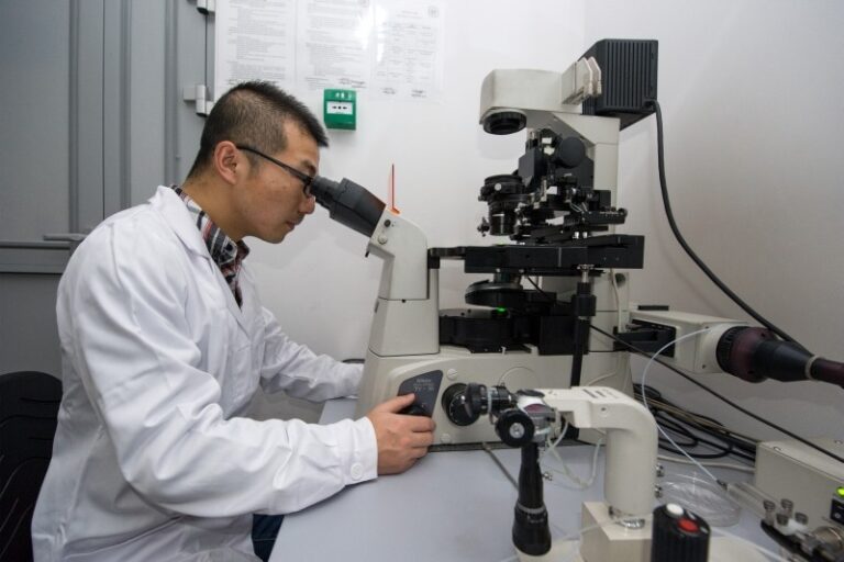
Researchers from the Institute of Physical Chemistry of the Polish Academy of Sciences have demonstrated, using a super resolution microscopic technique, how to follow chemical reactions taking place in very small volumes. The method of analysis developed by the Warsaw physicists in collaboration with PicoQuant GmbH is the first to make it potentially possible to observe reactions not only inside living cells, but even within individual organelles, such as cell nuclei.
The chemical mechanisms responsible for the cell’s vital functions still conceal many secrets. There is no surprise in this: only recently have we had the tools to directly look at the chemical phenomena occurring in living cells. However, due to continuing technical limitations, to this day we do not have, for example, such basic knowledge as that about the equilibrium constant values of chemical reactions in cells. In other words, we still do not know how much of a chemical involved in a given reaction in the cell is in an already reacted form and how much is in an unreacted form. The hitherto adversities have been overcome by a group of researchers from the Institute of Physical Chemistry of the Polish Academy of Sciences (IPC PAS) in Warsaw. In collaboration with the Berlin-based company PicoQuant GmbH, they have developed and demonstrated a modification of one of the most modern microscopic techniques: super resolution fluorescence correlation spectroscopy.
“We have been dealing with chemical reactions in cells for a long time. For example, in 2013, we determined the diffusion coefficients of all the proteins in the Escherichia coli bacterium, thanks to which it became possible to determine the rate of reactions taking place with their participation. Here we were interested in a similar issue, but with regard to the situation when we have low concentrations of reagents,” says Prof. Robert Holyst (IPC PAS) and continues: “Biological reactions are generally reversible and, where they occur, a certain dynamic equilibrium is usually created between the amount of reacted and unreacted substances. In our attempts to determine the equilibrium constants for various reactions in cells, we looked to super resolution fluorescence correlation spectroscopy. And here we came across an interesting technical problem whose solution opened up new possibilities for us in the study of the chemistry of life.”
There are many varieties of microscopy, including those with such phenomenal resolutions that it is possible to see individual atoms. However, when observing cells, optical microscopy remains unbeatable due to its low invasiveness and the ability to visualize the spatial structure of living organisms. For a long time, its basic disadvantage was its relatively poor resolution: fundamental physical constraints (diffraction) make it impossible to distinguish details smaller than about 200 nanometres by standard optical techniques.
One type of optical microscopy is fluorescence microscopy. It involves introducing a fluorescent dye into the sites of the biological sample being studied, and then scanning the sample with a focused laser beam. The dye molecules that are in the focus are stimulated to glow. In 1994, Stefan W. Hell presented a method of exceeding the diffraction limit in fluorescence microscopy by means of STIMulated Emission Depletion (STED). STED requires an additional laser beam, resembling a doughnut in cross-section. Properly used, this beam extinguishes the external areas of the main focus of the laser beam and consequently reduces its size to values below the diffraction limit. With super resolution methods it is now possible to see spatial details of only 10 nm with a time resolution of up to microseconds.
A relatively young branch of optical microscopy is Fluorescence Correlation Spectroscopy (FCS), used to study motion of molecules. In super resolution varieties, the focus of the laser has a volume measured in tens of attolitres (one attolitre is a billionth of a billionth of a litre). The measurement involves measuring the light emitted by a fluorescent dye attached to the tested molecule, excited by a laser beam. Knowing the size of the focus and the duration of fluorescence, and with the assistance of the appropriate theoretical models, it is possible to quite accurately determine the velocity of even individual molecules.
“For some time it has been known that while super resolution FCS microscopy works well when observing molecules moving in two dimensions, e.g. in lipid membranes, it fails in observations in volumes. Diffusion times, determined on the basis of measurements in 3D, could differ from the predictions from measurements in 2D by an order of magnitude or even more. After a few months of research it became clear to us that for these discrepancies were due to the excessively simplified manner of determining the spatial size of the focus,” says Dr. Krzysztof Sozanski (IPC PAS).
On the basis of their own theoretical analyses and experiences, the Warsaw researchers, funded by MAESTRO grant from the Polish National Science Centre and ERA Chairs grant from the European Horizon 2020 programme, constructed a new, universal theoretical model introducing a correction of the spatial shape of the focus and taking into account its impact on the measured signal-to-noise ratio. The correctness of the model was initially verified in measurements of the diffusion rate of various fluorescent probes in solutions.
“We also carried out more advanced experiments. For example, we studied a reversible reaction in which the dye molecules attached themselves to micelles and then detached themselves after some time. The system, composed of relatively large balls of surfactant molecules reacting with the molecules of dye, reflected conditions characteristic of biological structures,” says PhD student Xuzhu Zhang (IPC PAS). The measurements were not simple. If the molecules of both reactants were moving slowly, when passing through the focus the dye could repeatedly merge/disconnect with/from the micelles and the emitted light would be averaged. But there could also be a variant of the other extreme: the connection and disconnection reactions could run so slowly that during the transition through the focus there would be no change in the relationship between the reagents – then there would be no averaging. “Our model takes into consideration not only both of the extreme cases, but also all the intermediate ones. And with the knowledge at our disposal about the actual size of the focus, we are able to change its size and experimentally examine all the cases required by the model both in the same chemical system and on the same equipment,” emphasizes Zhang.
An important feature of the analytical method developed at the IPC PAS is the fact that no changes in apparatus are needed for its application. After appropriate adaptation, the method can be used to more accurately interpret data recorded by FCS-ready STED microscopes already in production.
This press release was prepared with funds from the European ERA Chairs grant under the Horizon 2020 programme.
Author: dr Krzysztof Sozański
Contact: ksozanski@ichf.edu.pl